
Melamine Sound Absorbing Foam Properties And Applications
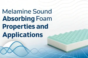
Tech Blog Melamine Sound Absorbing Foam Properties and Applications Melamine sound-absorbing foam is a high-performance open-cell foam with outstanding sound absorption, thermal insulation, flame-retardant properties,